Skin Anatomy 101
Our skin is the largest organ of the human body that covers the outside of the body. One inch of skin has approximately 19 million cells.
Skin has many functions. It is a barrier to the outside world protecting our internal organs from ultraviolet radiation, microscopic pathogens, physical or chemical injury and heat. It also helps us gather sensory information such as detecting temperature and touch. Generally, there are two types of skin: thin skin and thick skin. Thin skin is what covers the majority of our body. It contains hair follicles, sweat glands, nerves, blood vessels, and oil glands. Thick skin has the same layers as thin skin, but it contains an extra layer in the epidermis and is found only on the soles of the feet and palms of the hands.
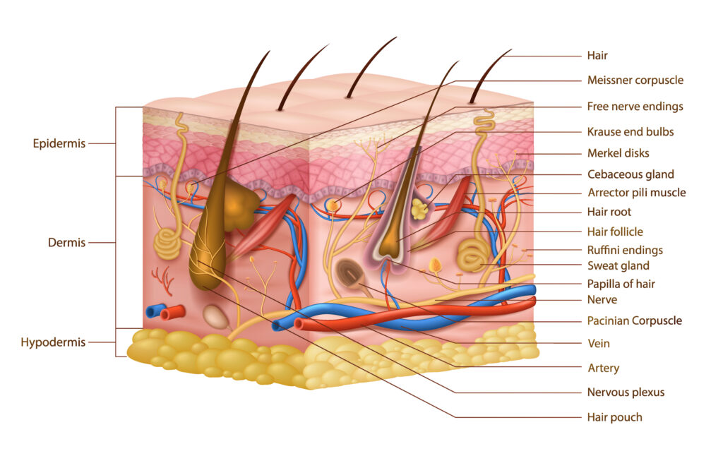
Skin anatomy is composed of the three main layers: the epidermis, the dermis, and the hypodermis. Each layer has its own function and many specialized structures which work together to protect the body from the outside world.
The Epidermis
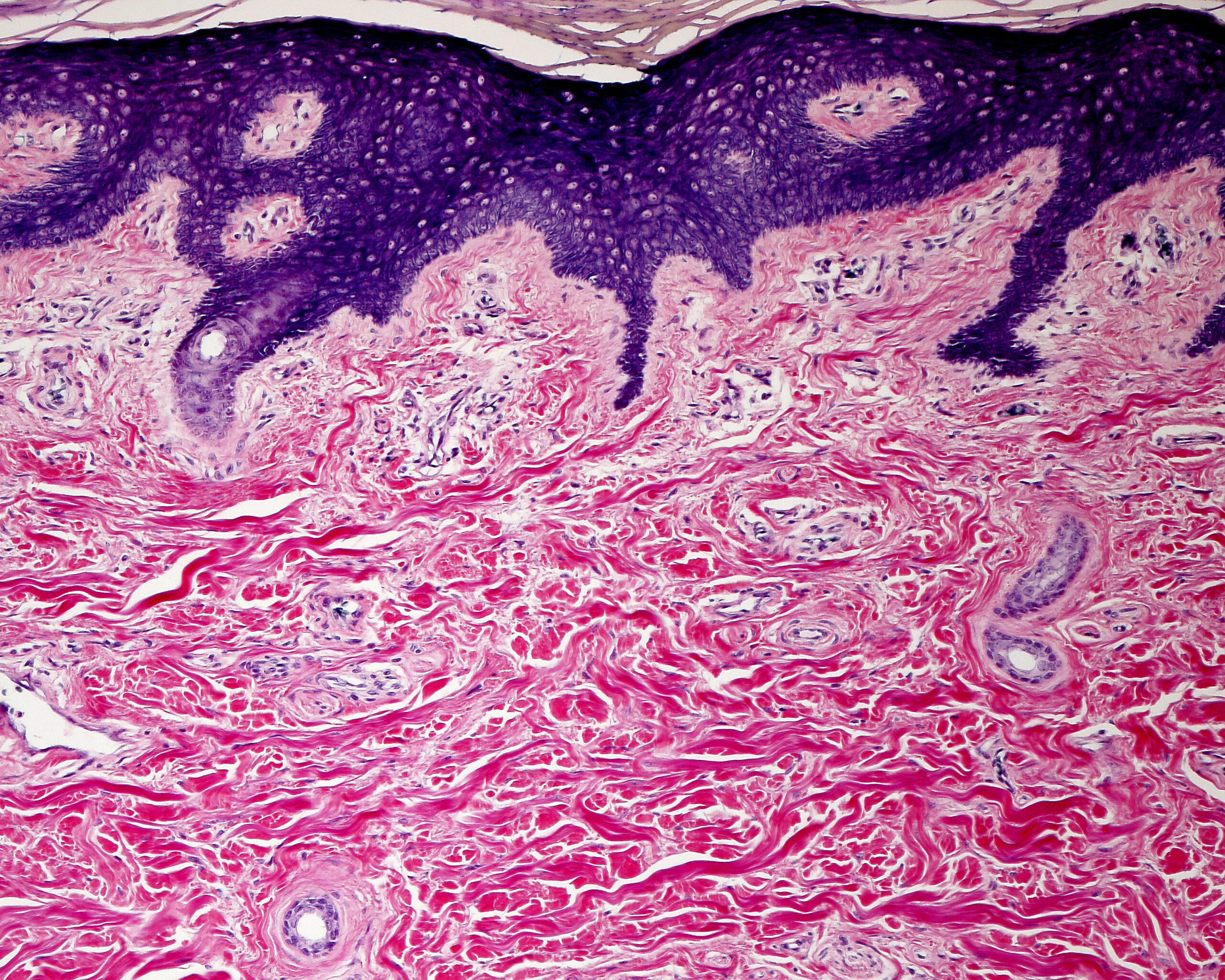
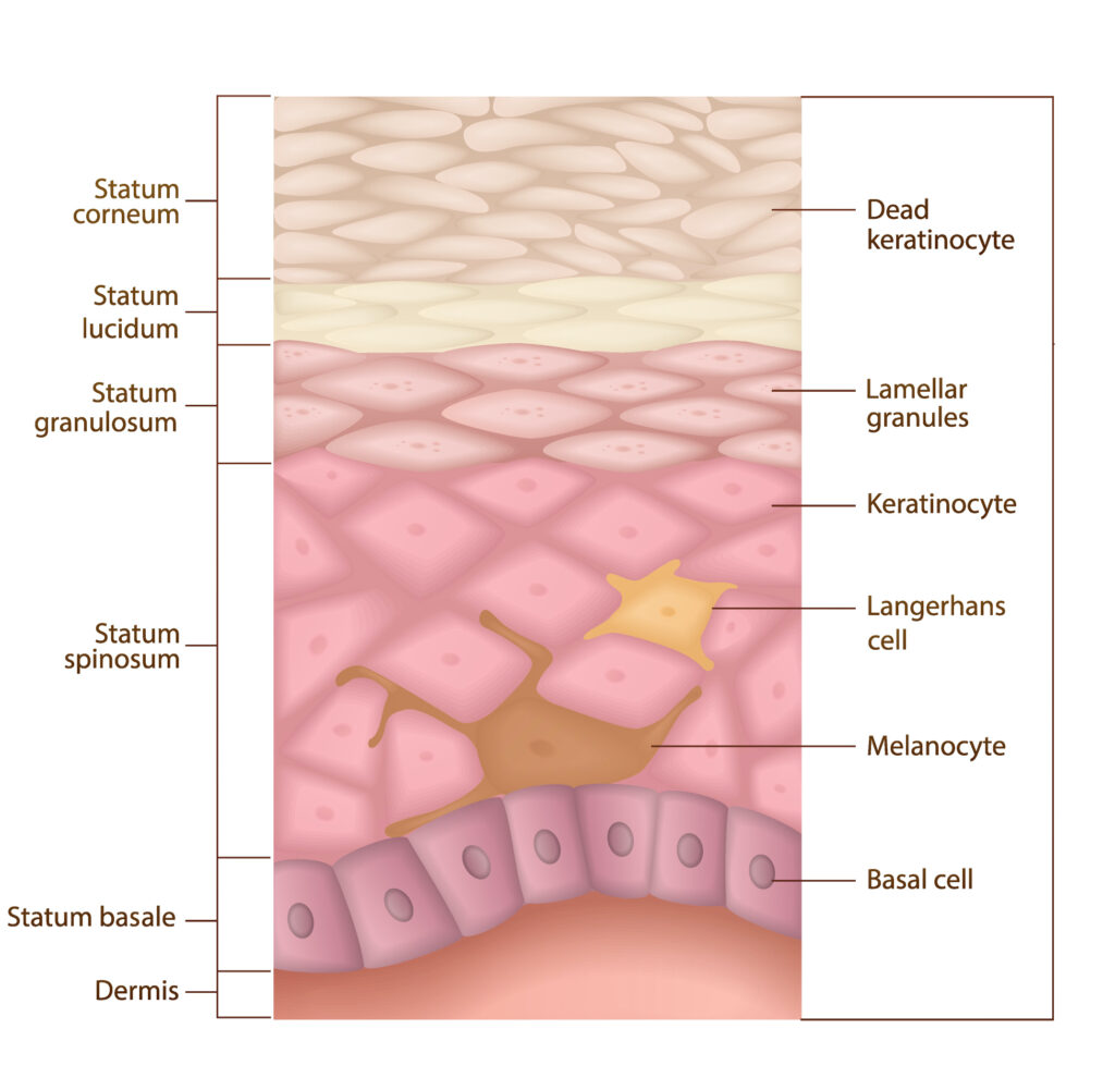
The most superficial layer and one we see easily with our eyes is the epidermis. If we were to zoom into the epidermis on a microscope we will see that it is actually made of five layers. It is the thinnest layer of the three main layers. In the diagram below you will see that the top layer of the epidermis is called the stratum cornuem and the bottom layer is the stratum basale.
Stratum Basale: The stratum basale the innermost or deepest layer of the epidermis where the cells multiply and undergo mitosis pushing older cells to the surface. This one row of cells look like columns or cubes lining side by side, and are constantly making keratinocytes, which is the major cell of the skin. It takes about 30 days for new cells formed in the stratum basale to travel upward towards the surface of our skin to get shed and replaced. The stratum basale contains melanocytes, which produce melanin to give skin its color and protect against UV radiation.
Stratum Spinosum: The stratum spinosum, also known as the prickle cell layer, gets its name from the spiny appearance of the keratinocytes when viewed under a microscope. This layer is approximately 5-10 cells thick and contains Langerhans cells, which play a role in the immune response. The stratum spinosum is responsible for:
- Support: Provides strength and flexibility to the skin.
- Immune Defense: Langerhans cells detect and fight off foreign invaders.
Stratum Granulosum: The stratum granulosum, or the granular layer, is where the keratinocytes begin to die and form a water-resistant barrier. This layer contains keratohyalin granules, which are essential for forming keratin in the upper layers. Functions of the stratum granulosum include:
- Waterproofing: Helps to create a lipid barrier, preventing water loss and entry.
- Strengthening: Contributes to the structural integrity of the skin.
Stratum Lucidium: The stratum lucidum is a thin, transparent layer found only in certain parts of the body, such as the palms of the hands and the soles of the feet. This layer is composed of dead keratinocytes that have lost their nuclei and are packed with a clear protein called eleidin. Its primary role is to provide an extra barrier to areas subject to friction and pressure.
Stratum Corneum: The stratum corneum is the topmost layer of the epidermis, and it's what we see when we look at our skin. This layer is composed of dead, flattened keratinocytes that are constantly shed and replaced. Within this layer, the dead keratinocytes secrete defensins which are part of our first immune defense. The primary functions of the stratum corneum include:
- Protection: Acts as a barrier against environmental damage and pathogens.
- Hydration: Retains moisture to prevent the skin from becoming dry and brittle.
- Regulation: Helps control the permeability of the skin, balancing the loss of water and electrolytes.
The Dermis
The dermis makes up 90% of the skin and is predominantly composed of collagen and elastin fibers, which provide the skin with strength, elasticity, and resilience. This middle layer contains the adjunctive structures such as nerves, hair follicles, sweat glands, collagen, oil glands and blood vessels. The sebaceous glands located next to the hair follicle create sebum that travels to the surface of the skin coating the skin with lipids and natural nourishment for protection. Sweat glands regulate temperate through evaporation and cooling. Arrector pilli muscles pull on the epidermis and hair follicle creating goose bumps when cold. The dermis plays a key role in wound healing and scar formation, making it a focal point in treatments aimed at anti-aging and skin rejuvenation.
The Hypodermis
This bottom layer, also known as the subcutaneous layer, set deep in the skin is made up of fatty cells. The fat tissues provide cushioning to the muscles and bones when you fall or scrap yourself. It acts as an insulator to help regulate the body's temperature by releasing and retaining heat. The blood vessels in the hypodermis are larger than the ones in the dermis, connecting blood flow in the dermis to the rest of the body.
Why Understanding the skin structure Matters
The skin is a remarkable, multi-layered structure that plays a vital role in protecting our body and maintaining overall skin health. By understanding its layers and functions, we can better appreciate the complexity of our skin and take informed steps to care of it. This building block of knowledge helps in understanding various skin conditions, such as psoriasis, eczema, and skin cancer, which often originate in the epidermis. This knowledge is vital for developing effective skincare routines and treatments aimed at maintaining or restoring the skin's health. Whether you're a dermatology enthusiast or simply someone interested in skincare, knowing about the basic anatomy of the skin is the first step towards achieving healthy, radiant skin.


